|
You
are here: /main/research
expeditions/May 2006/Days
7-8 FFS
Days
7-8, Coral genetics at the small scale
Morning of May 28, 2006
by
Dr. Malia Rivera
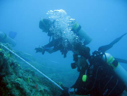
The
coral team surveying Porites lobata for fine scale genetic analyses.
Photo by Malia Rivera.
For
two days at French
Frigate Shoals the coral team has been searching for
a certain kind of coral reef patch. Small enough to be
practical for looking at every coral colony on it, but
large enough that adequate numbers of colonies would be
available for sampling,
allowing for a statistically valid investigation. After surveying
several sites, the coral team, made up of up to eight
divers at a time, finally found one that fit the bill.
About the size of a basketball court, a small
patch reef off Tern Island was home to just about the perfect
number of colonies of the Lobe coral, Porites lobata.
Like most species of corals,
the Lobe coral propagates either through fragmentation, whereby newly formed colonies are genetic clones
of the parent, or through sexual reproduction, where each new colony is a product of a set of two
parents that each contributed genetic variation. With this in mind, Dr. Stephen Karl of HIMB is
attempting to characterize the genotypes of coral colonies within a single patch reef. Why? He responds
with total sincerity, Every time I swim over a reef I see all these differences between colonies of
the same species of coral. Different colors, different morphologies, some bleached or diseased, some not,
and there all right next to each other, he explains. And I cant help but wonder why. So, he is
going try and find out.
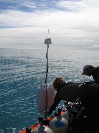 Steve marks the precise
location of every coral colony his team samples using a sophisticated GPS system called the GIS
Intelligent Buoy System, or GIBS for short. To do this, each of four buoys is placed about 400
meters away around the patch reef, equipped with a GPS unit and a hydrophone that communicates
with a diver-held acoustic transmitter and an onboard GPS unit. Triangulation allows for a
precise determination of each colony position, which is then automatically recorded onto a computer.
In addition to GPS locations, each colony is individually photographed to document any visible
variation, for
example in color, shape, or the presence of bleaching or disease. Steve marks the precise
location of every coral colony his team samples using a sophisticated GPS system called the GIS
Intelligent Buoy System, or GIBS for short. To do this, each of four buoys is placed about 400
meters away around the patch reef, equipped with a GPS unit and a hydrophone that communicates
with a diver-held acoustic transmitter and an onboard GPS unit. Triangulation allows for a
precise determination of each colony position, which is then automatically recorded onto a computer.
In addition to GPS locations, each colony is individually photographed to document any visible
variation, for
example in color, shape, or the presence of bleaching or disease.
Back in the lab, Steve will be taking each of the small coral samples and looking at
relatedness using a series of highly variable genetic markers known as microsatellites.
Because this type of DNA accumulates changes very quickly, they are particularly useful
for distinguishing relatively close genetic relationships. The more such microsatellites
markers used, the increased chance Steve will be able to distinguish between which
colonies are identical (and therefore clones), which are only distantly related, and
every level of relatedness in between. Using this kind of data, Steve can tell just how
many genetic individuals make up a reef. He can also then be compare the results
against the photographs to further investigate whether certain genetic types are
associated with different colors or morphologies, or more importantly, disease or
bleaching.
|